Identifying Oral Cysts and Growths: Pathology Care in Massachusetts
Massachusetts patients frequently arrive at the oral chair with a small riddle: a pain-free swelling in the jaw, a white patch under the tongue that does not rub out, a tooth that refuses to settle despite root canal therapy. Most do not come inquiring about oral cysts or tumors. They come for a cleansing or a crown, and we discover something that does not fit. The art and science of identifying the harmless from the dangerous lives at the crossway of scientific watchfulness, imaging, and tissue medical diagnosis. In our state, that work pulls in several specializeds under one roof, from Oral and Maxillofacial Pathology and Radiology to Surgery and Oral Medication, with support from Endodontics, Periodontics, Prosthodontics, and even Orthodontics and Dentofacial Orthopedics. When the handoff is smooth, clients get the answer quicker and treatment that appreciates both biology and function.
What counts as a cyst, what counts as a tumor
The words feel heavy, however they explain patterns of tissue growth. An oral cyst is a pathological cavity lined by epithelium, often filled with fluid or soft particles. Numerous cysts emerge from odontogenic tissues, the tooth-forming device. A tumor, by contrast, is a neoplasm: a clonal expansion of cells that can be benign or deadly. Cysts enlarge by fluid pressure or epithelial proliferation, while growths enlarge by cellular development. Clinically they can look similar. A rounded radiolucency around a tooth root might be a benign radicular cyst, an odontogenic keratocyst, or the early face of an ameloblastoma. All 3 can provide in the exact same years of life, in the same area of the mandible, with similar radiographs. That uncertainty is why tissue diagnosis remains the gold standard.
I typically inform clients that the mouth is generous with warning signs, however likewise generous with mimics. A mucous retention cyst on the lower lip looks obvious when you have seen a hundred of them. The first one you fulfill is less cooperative. The same logic applies to white and red patches on the mucosa. Leukoplakia is a medical descriptor, not a diagnosis. It can represent frictional keratosis, lichen planus, or a dysplastic process on the path to oral squamous cell cancer. The stakes vary immensely, so the procedure matters.
How problems expose themselves in the chair
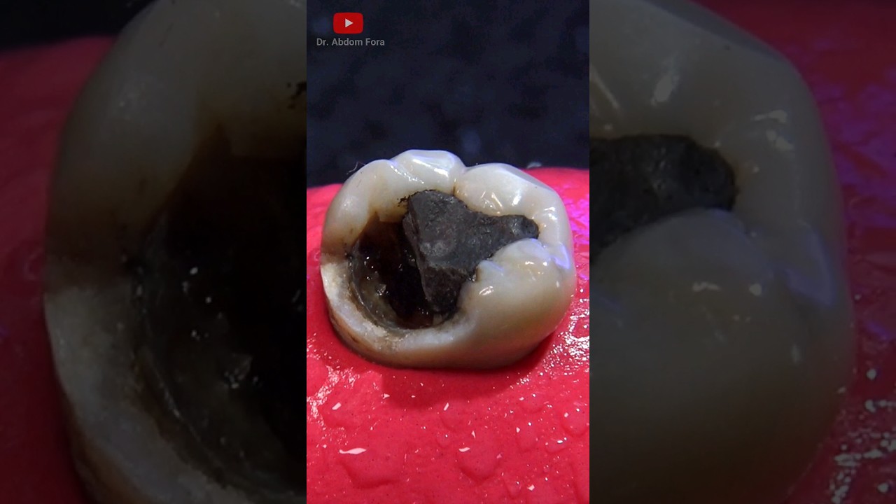
The most typical course to a cyst or growth medical diagnosis begins with a routine examination. Dental professionals identify the peaceful outliers. A unilocular radiolucency near the apex of a previously treated tooth can be a relentless periapical cyst. A well-corticated, scalloped sore interdigitating in between roots, centered in the mandible between the canine and premolar region, may be a simple bone cyst. A teen with a slowly expanding posterior mandibular swelling that has actually displaced unerupted molars might be harboring a dentigerous cyst. And a unilocular lesion that appears to hug the crown of an impacted tooth can either be a dentigerous cyst or the less courteous cousin, a unicystic ameloblastoma.
Soft tissue clues require equally steady attention. A patient complains of a sore area under the denture flange that has actually thickened in time. Fibroma from chronic trauma is likely, however verrucous hyperplasia and early cancer can embrace similar disguises when tobacco belongs to the history. An ulcer that continues longer than 2 weeks should have the self-respect of a medical diagnosis. Pigmented sores, particularly if asymmetrical or altering, need to be documented, determined, and frequently biopsied. The margin for mistake is thin around the lateral tongue and floor of mouth, where malignant improvement is more common and where growths can conceal in plain sight.
Pain is not a dependable narrator. Cysts and numerous benign tumors are pain-free till they are big. Orofacial Pain professionals see the opposite of the coin: neuropathic pain masquerading as odontogenic illness, or vice versa. When a mystery toothache does not fit the script, collective review prevents the double risks of overtreatment and delay.
The role of imaging and Oral and Maxillofacial Radiology
Radiographs fine-tune, they hardly ever complete. A knowledgeable Oral and Maxillofacial Radiology team reads the subtleties of border definition, internal structure, and effect on surrounding structures. They ask whether a lesion is unilocular or multilocular, whether it triggers root resorption or tooth displacement, whether it broadens or perforates cortical plates, and whether the mandibular canal is displaced inferiorly or superimposed.
For cystic sores, breathtaking radiographs and periapicals are typically sufficient to define size and relation to teeth. Cone beam CT adds important detail when surgical treatment is likely or when the sore abuts important structures like the inferior alveolar nerve or maxillary sinus. MRI plays a restricted however meaningful role for soft tissue masses, vascular abnormalities, and marrow infiltration. In a practice month, we may send out a handful of cases for MRI, usually when a mass in the tongue or flooring of mouth requires better soft tissue contrast or when a salivary gland tumor is suspected.
Patterns matter. A multilocular "soap bubble" look in the posterior mandible nudges the differential toward ameloblastoma or odontogenic myxoma. A well-circumscribed, corticated radiolucency connected at the cementoenamel junction of an affected tooth recommends a dentigerous cyst. A radiolucency at the pinnacle of a non-vital tooth highly favors a periapical cyst or granuloma. But even the most book image can not replace histology. Keratocystic sores can present as unilocular and harmless, yet act strongly with satellite cysts and greater recurrence.
Oral and Maxillofacial Pathology: the response is in the slide
Specimens do not speak until the pathologist provides a voice. Oral and Maxillofacial Pathology brings that accuracy. Biopsy choice is part science, part logistics. Excisional biopsy is ideal for little, well-circumscribed soft tissue sores that can be gotten rid of completely without morbidity. Incisional biopsy suits large sores, locations with high suspicion for malignancy, or sites where complete excision would run the risk of function.
On the bench, hematoxylin and eosin staining stays the workhorse. Special discolorations and immunohistochemistry help distinguish spindle cell tumors, round cell tumors, and poorly differentiated cancers. Molecular studies sometimes deal with rare odontogenic tumors or salivary neoplasms with overlapping histology. In practice, a lot of regular oral lesions yield a diagnosis from standard histology within a week. Deadly cases get sped up reporting and a phone call.
It deserves mentioning clearly: no clinician must feel pressure to "guess right" when a sore is relentless, atypical, or positioned in a high-risk site. Sending tissue to pathology is not an admission of unpredictability. It is the requirement of care.
When dentistry becomes group sport
The finest outcomes show up when specializeds align early. Oral Medication often anchors that process, triaging mucosal disease, immune-mediated conditions, and undiagnosed pain. Endodontics helps identify relentless apical periodontitis from cystic modification and manages teeth we can keep. Periodontics examines lateral periodontal cysts, intrabony flaws that simulate cysts, and the soft tissue architecture that surgery will need to respect afterward. Oral and Maxillofacial Surgical treatment provides biopsy and definitive enucleation, marsupialization, resection, and restoration. Prosthodontics expects how to restore lost tissue and teeth, whether with fixed prostheses, overdentures, or implant-supported services. Orthodontics and Dentofacial Orthopedics joins when tooth movement is part of rehabilitation or when impacted teeth are knotted with cysts. In complicated cases, Oral Anesthesiology makes outpatient surgical treatment safe for clients with medical complexity, dental stress and anxiety, or procedures that would be dragged out under regional anesthesia alone. Oral Public Health enters play when gain access to and prevention are the challenge, not the surgery.
A teenager in Worcester with a big mandibular dentigerous cyst gained from this choreography. After imaging and biopsy, we marsupialized the cyst to decompress it, protected the inferior alveolar nerve, and protected the establishing molars. Over six months, the cavity diminished by more than half. Later, we enucleated the residual lining, grafted the problem with a particulate bone replacement, and coordinated with Orthodontics to guide eruption. Final count: natural teeth protected, no paresthesia, and a jaw that grew typically. The option, a more aggressive early surgical treatment, may have gotten rid of the tooth buds and produced a larger defect to reconstruct. The option was not about bravery. It had to do with biology and timing.
Massachusetts pathways: where patients go into the system
Patients in Massachusetts move through numerous doors: private practices, neighborhood health centers, healthcare facility dental centers, and academic centers. The channel matters because it defines what can be done in-house. Community centers, supported by Dental Public Health initiatives, frequently serve clients who are uninsured or underinsured. They may do not have CBCT on website or easy access to sedation. Their strength depends on detection and recommendation. A small sample sent out to pathology with an excellent history and picture often reduces the journey more than a lots impressions or duplicated x-rays.
Hospital-based clinics, including the dental services at academic medical centers, can finish the complete arc from imaging to surgical treatment to prosthetic rehab. For malignant growths, head and neck oncology teams coordinate neck dissection, microvascular restoration, and adjuvant treatment. When a benign however aggressive odontogenic growth requires segmental resection, these teams can provide fibula flap reconstruction and later implant-supported Prosthodontics. That is not most clients, however it is great to know the ladder exists.
In private practice, the best path is a network. Know your closest Oral and Maxillofacial Radiology service for CBCT checks out, your chosen Oral and Maxillofacial Surgical treatment team for biopsies, and an Oral Medicine coworker for vexing mucosal disease. Massachusetts licensing and recommendation patterns make cooperation uncomplicated. Patients value clear descriptions and a plan that feels intentional.
Common cysts and tumors you will actually see
Names collect quickly in textbooks. In day-to-day practice, a narrower group represent most findings.
Periapical (radicular) cysts follow non-vital teeth and chronic inflammation at the peak. They present as round or ovoid radiolucencies with corticated borders. Endodontic treatment solves many, but some persist as real cysts. Persistent sores beyond 6 to 12 months after quality root canal therapy deserve re-evaluation and often apical surgical treatment with enucleation. The diagnosis is excellent, though large sores may need bone grafting to support the site.
Dentigerous cysts connect to the crown of an unerupted tooth, frequently mandibular 3rd molars and maxillary dogs. They can grow silently, displacing teeth, thinning cortex, and in some cases expanding into the maxillary sinus. Enucleation with elimination of the included tooth is basic. In more youthful patients, cautious decompression can conserve a tooth with high aesthetic value, like a maxillary canine, when integrated with later orthodontic traction.
Odontogenic keratocysts, now frequently identified keratocystic odontogenic tumors in some categories, have a track record for reoccurrence since of their friable lining and satellite cysts. They can be unilocular or multilocular, typically in the posterior mandible. Treatment balances reoccurrence danger and morbidity: enucleation with peripheral ostectomy prevails. Some centers use accessories like Carnoy option, though that option depends on distance to the inferior alveolar nerve and evolving proof. Follow-up spans years, not months.
Ameloblastoma is a benign growth with deadly behavior towards bone. It pumps up the jaw and resorbs roots, seldom metastasizes, yet repeats if not fully excised. Small unicystic variants abutting an affected tooth in some cases respond to enucleation, specifically when validated as intraluminal. Strong or multicystic ameloblastomas usually need resection with margins. Reconstruction varieties from titanium plates to vascularized bone flaps. The choice depends upon place, size, and client priorities. A patient in their thirties with a posterior mandibular ameloblastoma will live longest with a durable solution that protects the inferior border and the occlusion, even if it demands more up front.
Salivary gland tumors occupy the lips, taste buds, and parotid region. Pleomorphic adenoma is the timeless benign tumor of the taste buds, firm and slow-growing. Excision with a margin prevents reoccurrence. Mucoepidermoid carcinoma appears in small salivary glands more often than a lot of expect. Biopsy guides management, and grading shapes the requirement for broader resection and possible neck examination. When a mass feels fixed or ulcerated, or when paresthesia accompanies growth, escalate quickly to an Oral and Maxillofacial Surgical treatment or head and neck oncology team.
Mucoceles and ranulas, common and mercifully benign, still benefit from correct strategy. Lower lip mucoceles deal with finest with excision of the lesion and associated minor glands, not mere drain. Ranulas in the flooring of mouth often trace back to the sublingual gland. Marsupialization can help in small cases, but elimination of the sublingual gland addresses the source and minimizes recurrence, especially for plunging ranulas that extend into the neck.
Biopsy and anesthesia choices that make a difference
Small procedures are much easier on patients when you match anesthesia to character and history. Many soft tissue biopsies are successful with local anesthesia and simple suturing. For patients with serious oral stress and anxiety, neurodivergent clients, or those needing bilateral or multiple biopsies, Oral Anesthesiology broadens alternatives. Oral sedation can cover straightforward cases, but intravenous sedation provides a foreseeable timeline and a safer titration for longer treatments. In Massachusetts, outpatient sedation needs appropriate allowing, tracking, and personnel training. Well-run practices document preoperative evaluation, air passage assessment, ASA classification, and clear discharge requirements. The point is not to sedate everybody. It is to remove gain access to barriers for those who would otherwise prevent care.
Where avoidance fits, and where it does not
You can not avoid all cysts. Many emerge from developmental tissues and hereditary predisposition. You can, however, avoid the long tail of damage with early detection. That starts with consistent soft tissue exams. It continues with sharp photographs, measurements, and accurate charting. Cigarette smokers and heavy alcohol users bring higher danger for malignant change of oral possibly deadly disorders. Counseling works best when it is specific and backed by recommendation to cessation assistance. Dental Public Health programs in Massachusetts frequently offer resources and quitlines that clinicians can hand to clients in the moment.
Education is not scolding. A client who comprehends what we saw and why we care is more likely to return for the re-evaluation in two weeks or to accept a biopsy. An easy phrase helps: this area does not act like normal tissue, and I do not wish to guess. Let us get the facts.
After surgical treatment: bone, teeth, and function
Removing a cyst or growth creates an area. What we finish with that area identifies how rapidly the patient go back to normal life. Small flaws in the mandible and maxilla often fill with bone in time, specifically in younger clients. When walls are thin or the problem is big, particulate grafts or membranes support the site. Periodontics often guides these choices when surrounding teeth require foreseeable assistance. When lots of teeth are lost in a resection, Prosthodontics maps completion game. An implant-supported prosthesis is not a high-end after significant jaw surgery. It is the anchor for speech, chewing, and confidence.
Timing matters. Placing implants at the time of cosmetic surgery matches particular flap reconstructions and patients with travel burdens. In others, delayed placement after graft consolidation lowers risk. Radiation treatment for deadly disease changes the calculus, increasing the threat of osteoradionecrosis. Those cases require multidisciplinary planning and typically hyperbaric oxygen only when evidence and risk profile validate it. No single rule covers all.
Children, households, and growth
Pediatric Dentistry brings a various lens. In children, sores interact with development centers, tooth buds, and airway. Sedation choices adapt. Behavior assistance and adult education become main. A cyst that would be enucleated in a grownup may be decompressed in a kid to preserve tooth buds and minimize structural impact. Orthodontics and Dentofacial Orthopedics frequently joins quicker, not later on, to direct eruption courses and prevent secondary malocclusions. Moms and dads appreciate concrete timelines: weeks for decompression and dressing modifications, months for shrinkage, a year for last surgery and eruption guidance. Vague strategies lose families. Specificity builds trust.
When pain is the problem, not the lesion
Not every radiolucency discusses pain. Orofacial Pain experts advise us that consistent burning, electrical shocks, or hurting without provocation might show neuropathic processes like trigeminal neuralgia or consistent idiopathic facial pain. Alternatively, a neuroma or an intraosseous lesion can present as pain alone in a minority of cases. The discipline here is to prevent heroic oral treatments when the discomfort story fits a nerve origin. Imaging that fails to associate with signs ought to trigger a time out and reconsideration, not more drilling.
Practical cues for daily practice
Here is a short set of hints that clinicians throughout Massachusetts have discovered useful when navigating suspicious sores:
- Any ulcer lasting longer than two weeks without an obvious cause deserves a biopsy or instant referral.
- A radiolucency at a non-vital tooth that does not diminish within 6 to 12 months after well-executed Endodontics needs re-evaluation, and typically surgical management with histology.
- White or red spots on high-risk mucosa, particularly the lateral tongue, flooring of mouth, and soft taste buds, are not watch-and-wait zones; file, photograph, and biopsy.
- Rapidly growing swellings, paresthesia, or spontaneous bleeding shift cases out of routine paths and into immediate assessment with Oral and Maxillofacial Surgery or Oral Medicine.
- Patients with risk aspects such as tobacco, alcohol, or a history of head and neck cancer gain from shorter recall intervals and precise soft tissue exams.
The public health layer: gain access to and equity
Massachusetts succeeds compared to many states on dental access, but gaps continue. Immigrants, elders on fixed incomes, and rural locals can deal with delays for innovative imaging or expert visits. Oral Public Health programs push upstream: training medical care and school nurses to acknowledge oral red flags, moneying mobile centers that can triage and refer, and building teledentistry links so a suspicious sore in Pittsfield can be reviewed by an Oral and Maxillofacial Pathology team in Boston the same day. These efforts do not change care. They shorten the range to it.
One little step worth embracing in every office is a photograph procedure. An easy intraoral camera image of a lesion, conserved with date and measurement, makes teleconsultation significant. The difference between "white spot on tongue" and a high-resolution image that reveals borders and texture can identify whether a client is seen next week or next month.
Risk, reoccurrence, and the long view
Benign does not always indicate short. Odontogenic keratocysts can leading dentist in Boston repeat years later, in some cases as new lesions in different quadrants, particularly in syndromic contexts like nevoid basal cell cancer syndrome. Ameloblastoma can recur if margins were close or if the variant was mischaracterized. Even common mucoceles can repeat when minor glands are not removed. Setting expectations safeguards everyone. Clients deserve a follow-up schedule customized to the biology of their lesion: yearly breathtaking radiographs for numerous years after a keratocyst, scientific checks every 3 to 6 months for mucosal dysplasia, and earlier visits when any brand-new symptom appears.
What good care seems like to patients
Patients keep in mind 3 things: whether someone took their concern seriously, whether they understood the strategy, and whether pain was managed. That is where professionalism programs. Use plain language. Prevent euphemisms. If the word growth applies, do not change it with "bump." If cancer is on the differential, say so carefully and describe the next steps. When the lesion is likely benign, describe why and what confirmation involves. Offer printed or digital guidelines that cover diet plan, bleeding control, and who to call after hours. For anxious patients, a quick walkthrough of the day of biopsy, consisting of Dental Anesthesiology choices when proper, lowers cancellations and enhances experience.
Why the information matter
Oral and Maxillofacial Pathology is not a world apart from day-to-day dentistry in Massachusetts. It is woven into the recalls, the emergency gos to, the ortho seek advice from where an impacted canine declines to budge, and the prosthodontic case where a ridge swelling appears under a new denture. The details of identification, imaging, and medical diagnosis are not scholastic difficulties. They are patient safeguards. When clinicians embrace a constant soft tissue test, preserve a low threshold for biopsy of consistent sores, work together early with Oral and Maxillofacial Radiology and Surgical treatment, and align rehab with Periodontics and Prosthodontics, patients get timely, total care. And when Dental Public Health broadens the front door, more patients get here before a small problem becomes a huge one.
Massachusetts has the clinicians and the infrastructure to provide that level of care. recommended dentist near me The next suspicious sore you discover is the right time to utilize it.